How 2 in 1 Bioplasm NLS work
- Russell
- June 06, 2025
- 381
- 0
- 0
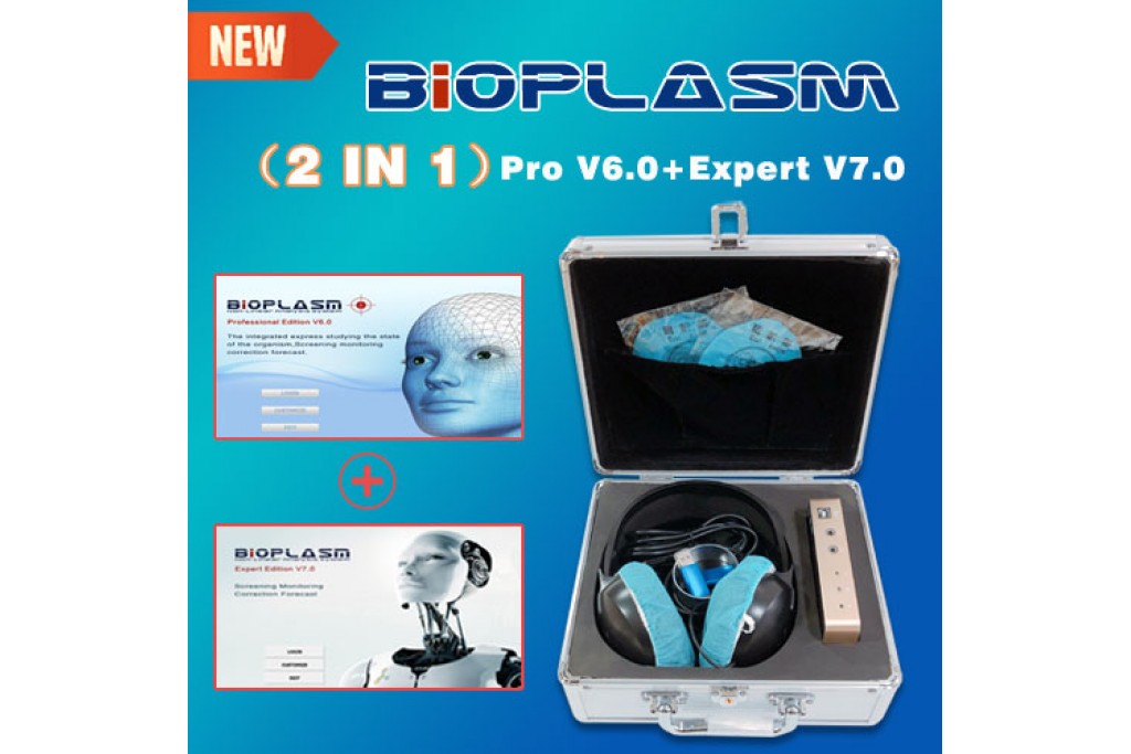
The device works on the principle of amplification of the iniciated signals with the disintegration of metastable structures. By influencing external electromagnetic fields the magnetic moments of the molecular currents in the centers of the cortex nerve cells are loosing their authentic orientation. This causes faulty adjustment of eddy structures of delocalized electrons which lead to formation of unstable metastable states. Disintegration of such state acts as iniciated signal. Expressed physically this device is system of the electronic oscillator which oscillates at appropriate wavelength of electromagnetic radiation. Its energy corresponds to the energy that degrades dominant bonds that maintain structural organization of biological objects in a good condition.The 2 in 1 Bioplasm NLS can cause a bioelectric aktivity of the brain cells so it is possible to selectively amplify signals in the background, which compared to static currents, are hard to detect. Information concerning specific temporal condition of organs and tissues are gathered on the basis of non-contact sensor that was developed using modern information technology and infrasonic circuits. This sensor reveals hardly detectable fluctuation of signals, which are filtered from sound fields and subsequently converted into a digital sequence where they are processed by a microprocessor and, using the interface cable, transferred to a computer.The development of a new generation of nonlinear computer scanners (metatrons), which make use of multidimensional virtual imaging of the object under investigation, has enabled to substantially enhance the effectiveness of the NLS-method and even expand its field of application, despite the competitiveness from MRT. The distinctive feature of multidimensional NLS imaging is an initially volumetric nature of scanning.
The data thus received are an integral array, which facilitates the reconstruction of multidimensional virtual images of anatomical structures of the object under investigation. In this connection the virtual NLS is widely used, especially for angiographic investigations with a three-dimensional reconstruction of vascular formations. The specific characteristic of the representation of virtual data by the Hunter system is simultaneous of surfaces of cavatus and extramural formations located outside the lumen of the cavity under examination (lymph nodes, vessels). The images that are received form a natural sequence of virtual NLS pictures.

